When this technology is used during liver surgery, it can help surgeons locate and reach tumors more efficiently.
Nowadays, the surgical intervention for treatment of liver tumours is planned based on preoperative images, typically CT or MR. These images provide a detailed representation of the patient’s anatomy and provides the ground information to plan the intervention. Unfortunately, these images are taken time before the operation, and thus, they do not show what is happening inside the body during the procedure.
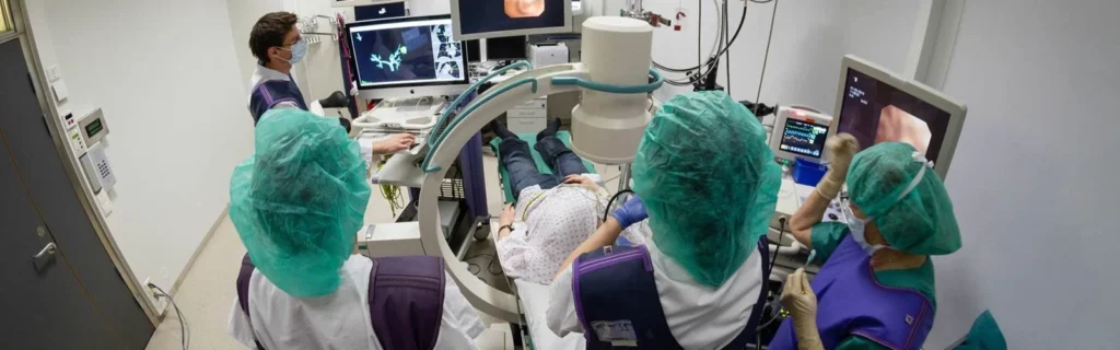
During the operation itself, the surgeons often use ultrasound to investigate the liver in real time. This provides updated information, though ultrasound can be difficult to interpret and do not always provide a view as good as CT or MR.
If preoperative detailed images are combined with real-time intraoperative ultrasound, the surgeons would be equipped with a detail rich and updated “map” of the liver during the intervention. This is technologically challenging; yet it would open the possibility for more precise surgical treatment.
SINTEF researchers work together with surgeons at St. Olavs hospital to make this technology a reality. With help of advanced software and navigation we can bring together different image modalities and display them in a way that helps surgeons to locate the tumours with higher precision. At the end of the day, we aim to enable safer, faster, and more precise interventions.
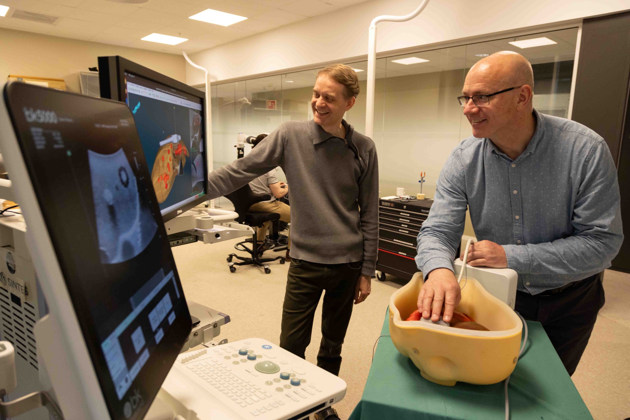
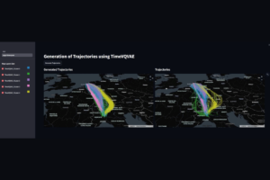
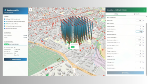
The technology combines CT images of the liver taken beforehand, with ultrasound images taken during the interventions. A 3D representation of the CT is shown on the screen.
Ole Vegard Solberg (left), research engineer at SINTEF Digital, and Torgrim Lie, research scientist at SINTEF Digital, bring together different technologies to navigate through a phantom of the liver.
With this pointer the researchers can mark where in the liver they are. This step is crucial to link the CT images of the liver to the real phantom which will be treated, an operation called registration. In a real operation, the surgeon would point the selected points outside of the patient’s body and not directly at the liver.
The CT image can be used as a “map” of the tumours inside the liver.
The technology is still under development and is currently used only in research projects. Nonetheless, it is based on known technology used in brain surgery. In this scenario, the conditions are more stable as the brain moves less during the intervention.
Through improvements in visualization and navigation during the operation, the project will contribute towards more precise surgery, better decision support, and lay the foundation for future solutions in image-guided and AI-assisted liver surgery.
The research is supported by the Ministry of Health and Care Services and the EU.
All photos: Mari Aftret Mørtvedt / SINTEF
Health Research in SINTEF Digital
Medical Technology
We develop medical technology that makes diagnostics, treatment and training safer, more precise and more accessible.
Our core expertise lies in ultrasound, image-guided intervensions, medical software and simulation for the benefit of patients, clinicians and society.


Comments
No comments yet. Be the first to comment!